Our sense organs are very important, and the Eye, amongst all, cannot be overruled. The ability of the eyes being able to utilize the surrounding light to get visuals captures which is interpreted by the brain to help our other part of the body understand and function accordingly is one reason why it cannot be overruled. The eye is a combination of several intricate parts working together to give proper sight. Since the eye is responsible for over 80% of our expressions as well as reading the expressions of others, then a defect in the eye can be very harmful to our daily activities as well as to the brain when interpreting images. In this post, I will be carefully looking at Strabismus, a defect in the eye, but before then, we will have to do a very little dive into the anatomy of the eye.
Hey, aren't you tired of reading in layman terms because I am tired of writing in such way, but we need to make this scientific, readable, and enjoyable. Awhn! so let's dive in, keeping it simple.
The Anatomy of the Sight Machine
I will be explaining the Eye anatomy, starting with the Orbit, which is the body cavity that covers the eyeball, the muscles of the eyes, and the blood vessels. [I will be coming back to this, but let's quickly dive into the eye itself].
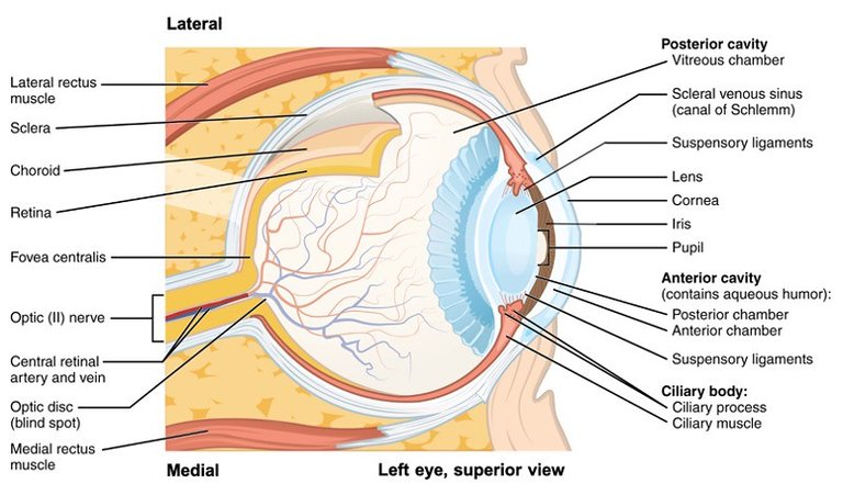
In explaining the anatomy of the eyeball, I will start with the Eye Tunics, and there are three tunics of the eye. The Fibrous tunic, the Vascular Tunic, and the Neuroectodermal/sensory tunic.
The Fibrous tunic is made up of the Sclera and the Cornea. The Sclera is the outer part of the eyes, made up of three layers which are the Episclera (have a dense fibrous), the Sclera (made up of collagen fibrous), and the Lamina Fusca (which is pigmented). Right in front of the Sclera is the Cornea.
The Cornea is a dome shaped, transparent part of the Tunic, projecting outwards. It is made up of 5 layers; the Epithelial layer (outer layer), Bowman's membrane, Corneal Stroma Layer, Descemet's Membrane, and the Endothelial Layer. The cornea allows for the passage of light.
The Vascular Tunic (uvea) is the middle layer surrounding the eye. It is made up of the choroid, Iris, and the Ciliary body (ciliary muscle, Ciliary zonular, Ciliary processes).
The Iris is a ring-shaped membrane, behind the cornea, that hold and control the pupil. The iris is made up of two different muscles called the Dilator pupillae (which dilates the pupil), and the Sphincter pupillae (which constricts the pupil). It is responsible for the color of the eyes as a result of pigments in the iris.
The Ciliary muscle which is connected to the lens of the eye, extends from the scleral spur, and is important in changing the shape of the lens in the process called accommodation reflex, which helps in controlling light penetration. Another part of the Ciliary body is the Ciliary Zonular which is a mesh fiber that serves as an anchor, holding the lens firm. When the Ciliary muscles contract, the zonular becomes loose, allowing the lens to bulge, giving close vision. When the Ciliary muscles relax, the zonular becomes tight causing the lens to be flattened, and allowing for longer vision. Not forgetting the Ciliary processes which are small, protrusions in the ciliary body, which produces aqueous humor which is formed by the inward folding of the choroid. The aqueous humor moves through the pupil creating lubrication for the eyes, washing metabolic wastes, and drain to the canal of schlemm. Not forgetting one part of the vascular tunic which is the Choroid. The Choroid is a pigmented part of the Uvea between the retina and the sclera. The choroid supplies nutrient, regulates temperature, and absorb light rays for the retina.
The Lens (which is made up of the lens capsule, the lens fibers and the lens epithelium), is responsible for refracting light rays to the retina.
The Neuroectodermal/Sensory/Nervous Tunic which basically is made up of the Retina. The retina is made up of ten layers (which I won't be discussing in this post). The retina is responsible for converting the light rays that penetrates into the eyes to electrical signals, which are then sent to the brain via the optic nerves.
The Extraocular Muscles
Moving forward from explaining the Eyeball, I will be going back to the orbit, but at this time, I will be looking at the Extraocular muscle. The muscle is responsible for the movement of the eyeball and the superior/upper eyelid of the eye.
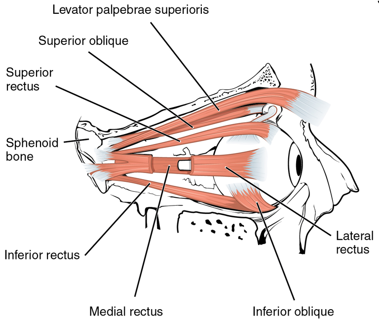
There are Six extraocular muscles, including the Levator palpebrae superioris. Four of these muscles are Recti Muscles, and the remaining two muscles are Oblique Muscles. The Recti Muscles includes; the Superior rectus, Inferior Rectus, Medial Rectus, and the Lateral Rectus. The Oblique muscles include; the superior oblique muscles, and the Inferior Oblique muscle.
The eyeball moves around three axises, which are the Horizontal axis, the vertical axis, and the anterior/posterior axis. The three axis allow the eyeball to move in different directions. When ever the Superior rectus contract, it pulls the eyeballs upwards. When the lateral rectus contract, it abducts the eyes, making it move laterally. When the Inferior rectus contracts, it causes a depression of the eyeball (causes the eyeball to look downwards). If the Medial Rectus Muscles contract, it pull the eyeball inwards/adducts the eyeball. When the Inferior Oblique muscle contracts, it pulls the eyeball upwards as well as laterally (this is because the muscle is attached to the Sclera). When the Superior oblique muscle contracts, it pulls the eyeball downwards. (Neuroscience Online).
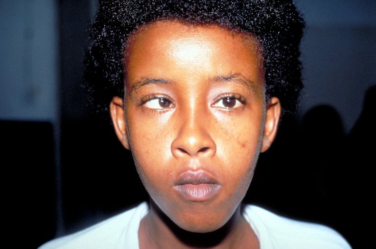
Strabismus (When an Extraocular Muscle is Faulty)
Just like the subtopic says, a Strabismus refers to a fault in the extraocular muscles of the eyes which causes maligned vision. When the eyes are normal, it is meant to move in a coordinated manner, thereby allowing the two eyeballs to move in the same direction.
According to a study published on the National Library of Medicine, the prevalence of Strabismus in the world ranges from 0.5% to 5%, and the prevalence in Ethiopia where the study was conducted can range from 1.5% to 17.9%. The research was conducted on 611 school children and 31 children, which is 5% of the children were confirmed to have Strabismus. Strabismus can be found in people of any age group (research gate).
Strabismus can be reffered to as Tropia when it is visible and Phoria when it is hidden. Pseudostrabismus is a very good class of strabismus that falls under Phoria. With Pseudostrabismus, the eyes appear misaligned, but they aren't.
Strabismus can occur as a result of a fracture from the Supranuclear structures, Oculomotor nuclei/nerves, and/or Extraocular muscles resulting from trauma. Extraocular muscle fracture can result in one or more muscles not functioning properly, leading to Strabismus. The fracture could be as a result of trauma from the muscle involvement in orbital wall (Ludwig 2002), or from the formation of edema or hematoma in the muscles thereby preventing it from functioning properly and getting fractured. According to Monte, trauma of the EOM can result in partial/complete loss of muscle function. At the point, the both eyeballs won't be miving in the same direction.
According to research by Ludwig 2002, msucles such as the inferior rectus, medial rectus, superior oblique, and inferior oblique, are prone to Orbital wall fracture. In the case of hematoma in the muscles, the strabismus is incomitant, since the muscles will still be functioning but not properly. The common EOM to experience this is the inferior rectus.
Another way Strabismus could occur is via the laceration of the Extraocular muscle. This could happen as a result of direct physical impact (accident, inserting sharp objects, hard blows and punches), thereby cutting the muscles.
When Diagnosing and Treating Strabismus, evaluation of the compromised muscle makes it easy to identify the degreee of damage the EOM has incured, as well as identify the condition of the sorrounding environment. treatment of Strabismus is often surgical, although some could be difficult to perform such as in cases of Laceration of the msucles . The surgery isn't always without complications in some cases. Complications could include; Orbital compartment syndrome, Permanent mydriasis, and Infraorbital nerve hypesthesia.
Conclusion
The eyes is a very delicate part of our body and should be cared for properly. In the case of Strabismus, it is very important that one identify eyeball movement disorder in children very fast, so as to help treat before it becomes a severe case as well as watch the toys being played with to prevent ocular injury. In cases where a person starts to experience double vision, difficulty in seeing, and cross-eye visions, visiting a doctor should be the next step. Self medication isn't accepted.
Image Reference
It is very important to get right away this problem
!1UP
Click this banner to join "The Cartel" discord server to know more.
You have received a 1UP from @gwajnberg!
@stem-curator, @neoxag-curator
And they will bring !PIZZA 🍕.
Learn more about our delegation service to earn daily rewards. Join the Cartel on Discord.
Thanks for your contribution to the STEMsocial community. Feel free to join us on discord to get to know the rest of us!
Please consider delegating to the @stemsocial account (85% of the curation rewards are returned).
Thanks for including @stemsocial as a beneficiary, which gives you stronger support.